Do you get hip pain is it fai!

Femoroacetabular Impingement (FAI) syndrome is a term used to describe a certain type of pain arising from the hip joint. Basically it is a situation where the bones of the hip abut each other resulting in pain in the bones themselves or pinching other material within the hip between these bones. It is related to the bony morphology (shape) of the hip joint and describes a range of things that can contribute to an imperfect fit of the ball (femoral head) within the socket (acetabulum).

Recent new guidelines (The Warwick Agreement on FAI) have identified 3 criteria that need to be present to be diagnosed with FAI syndrome. These include:

  1. Patient reported symptoms,
  2. Symptoms reproduced on clinical testing, and
  3. Changes on radiological imaging.

Without all three of these you cannot be diagnosed with FAI syndrome.

In a large proportion of people there may be bony changes on imaging without any symptoms. This is not FAI. It is important to remember that without symptoms these findings on imaging represent an individual’s normal bony morphology and does not necessarily need to be treated.

Symptoms?

The most common complaint of those with FAI syndrome is groin pain on the affected side, however this pain pattern can vary. Approximately 85% of those with FAI will have groin pain, 50% will have lateral (side) hip pain and around 5-10% will have posterior hip pain (bottom pain).

Usually these symptoms are reproduced with certain positions or movements – most commonly bending the hip up (flexion) and twisting movements. These may occur in everyday life in situations such as getting into a car or can occur during sporting or athletic activities.

Who is at risk?

Trying to determine who is at risk is a complex process. A large number of people will have deformities on imaging that do not correlate with pain thus imaging findings need to be taken with a grain of salt.

In saying that, those who have a history of hip problems, missing developmental milestones in childhood or a history of hip dysplasia (abnormal development) may be at an increased risk of FAI syndrome.

Treatment?

The evidence now suggests that an actual diagnosed FAI syndrome does not normally resolve on its own. If you have FAI syndrome there are broadly 3 treatment options:

  1. Conservative management – involves education on the condition especially around understanding positions which the hip is like to pinch in, activity modification to try to avoid these positions (usually only temporarily) and potentially pain relief using anti-inflammatories or cortisones injections.
  2. Physiotherapy rehabilitation – this will comprise of activity modification and education as above. In addition, a program of exercises looking at improving strength, range of motion and neuromuscular control of the hip would be implemented.
  3. Surgery – usually this is reserved for patients who have failed to respond to the above treatments. In broad terms, it aims to improve the structural “fit” of the ball in the socket.

If you think that you may have FAI, you should consult with one of our physiotherapists who have been trained to assess and treat this condition.

In addition, our Principal Physiotherapist, Brendan Limbrey, is a treating physiotherapist on a large international hip study (FASHIoN) for those with FAI which is looking at the outcomes gained from surgery compared with physiotherapy and conservative management.

Do you have a mortons foot

More specifically, a Morton’s foot is when the long bone in your foot connecting to your 2nd toe is longer than the long bone connecting to your big toe. You can recognise a Morton’s foot if the indent (gap) between your 1st and 2nd toe is lower than that of the indent (gap) between your 2nd and 3rd toe.

There is nothing abnormal with having a Morton’s foot as somewhere in the order of 20% of the population have them.

There are some subtle biomechanical diiferences between those who have a Morton’s foot and those that don’t. Having a Morton’s foot does not mean that you will have any weird or wonderful issues with your feet. In saying this though, we do know that having a Morton’s foot can place you at increased risk of:

  • plantar fasciitis
  • foot stress fractures
  • calluses under the ball of your foot
  • pain through the forefoot
  • neuromas (nerve pain)
  • hammer toes
  • having altered biomechanics which may cause ankle, knee or hip issues.

If you have a Morton’s foot and aren’t having any issue then there isn’t anything you need to do about it…you just know that your foot shape now has a name!

If you do have issues as outlined above then you should consult with a physiotherapist (or podiatrist) who understands how to assess and manage the conditions, taking into consideration your foot shape.

Cervicogenic Headaches

What are cervicogenic headaches?

Cervicogenic headaches describes a headache which is caused by irritation or dysfunction in your neck. These headaches can be caused by a number of issues within the neck including muscles, joints, nerves or ligaments in the neck. Typically, the area of concern is in the upper neck region. Each presentation is different and is typically a combination of issues to muscles, joints, nerves and ligaments in the upper neck.

How do these headaches present?

Common features of these headaches can be a constant dull ache. It will typically feel like it is coming from the base of your skull, the front of your face or behind your eyes. It can be felt on one or both sides of your head or even like a tight band around your head. As these types of headache originate from issues in your neck, it is common to feel some discomfort or restriction in your neck prior to headaches coming on. It is also common to feel that certain head and neck postures can influence your pain. It is also possible to feel dizzy, light headed or even nauseated when these headaches occur.

How can physiotherapy help cervicogenic headaches?

Firstly, it is important to recognise that there are numerous causes of headaches and it is important to determine whether issues in your neck are causing yours or whether it is being caused by other issues. A qualified physiotherapist can assess you and determine whether you are experiencing cervicogenic headaches and determine the factors influencing your pain. A full neck assessment and posture screen needs to be done so the best type of treatment can be tailored for you. Common treatments to assist in your acute cervicogenic headache symptoms can include posture education, joint mobilisations to target any restriction of movement in the neck joints and soft tissue release of the neck muscles. To ensure treatment is also focussed on the root cause a movement and strength assessment should be done. Strengthening of the deeper muscles of the neck along with muscles extending out to your shoulders can decrease the stress on your neck joints and help to improve your posture, in effort to prevent future headaches.

If you think you may be experiencing cervicogenic headaches, appointments with our physiotherapists can be made by phoning 8850 7770.

FIFA11+ Soccer warm up research update

Have you heard of the FIFA 11+?

If you are a soccer (or “real” football) player or coach, then we’re hoping that you have!

Whilst the soccer season might be starting the wrap-up, the following is super important and worth a read.

The FIFA 11+ is a warm-up program which has been specifically designed by the medical division of FIFA. It has been demonstrated to reduced injury rates and is applicable for all abilities.

If we get more specific, the FIFA 11+ is a neuromuscular training program. This means that it trains, develops and conditions the muscles and neural pathways in the body to ensure that your body knows how to appropriately deal with the situations which it is likely to encounter when playing soccer. The components therefore are very specific to soccer and regular completion of it is key.

Much of the research into the effectiveness of the FIFA 11+ in the past has been conducted with adolescent females. This is due to the fact that females have a higher rate of injury for certain key injuries, including serious knee injuries like Anterior Cruciate Ligament (ACL) ruptures. Whilst it may seem reasonable to be able to extrapolate the results of these studies to males, this does have its limitations, as one cannot simply presume that because something seems to happen in adolescent females that it would happen in mature males in the same way.

Good news for all you males out there! An article published in December of 2015 by Silvers-Granelli et al. looked at the efficacy of injury prevention using the FIFA 11+ on MALE collegiate soccer players.

What did they do?

65 teams of national level college soccer players were invited to participate in the study. These teams were randomised into either a control group or a the FIFA 11+ group. The FIFA 11+ group completed the FIFA 11+ program as a warm up 3 times a week. The control group performed no additional training on top of normal levels. This was performed over the course of one soccer season.

What were they looking for?

The study looked at:

  • How often the teams/players played & trained,
  • How many injuries occurred,
  • Compliance of the FIFA 11+ group using the FIFA 11+ program.

What did they find?

A study found a number of things including:

  • The FIFA 11+ group had significantly fewer injuries per teams compared to the control group,
  • The FIFA 11+ group had a lower number of injuries during both training & games compared to the control group,
  • There was a significant relationship identified with respect to utilisation and compliance of the FIFA 11+ program within the FIFA 11+ group. That being, the greater the compliance to the FIFA 11+ program, the lower the injury rate.
  • Injury rates in the FIFA 11+ group were significantly lower as well when accounted for injury type:
    • Control group reported 115 ankle injuries, whereas the FIFA 11+ group reported 59 ankle injuries;
    • Control group reported 102 knee injuries, whereas the FIFA 11+ group reported 34 knee injuries;
    • There were 16 ACL injuries in the control group compared to 3 ACL injuries in the FIFA 11+ group – representing a greater than 4x reduction in ACL injuries;
    • 55 hamstring injuries were reported in the control group compared to 16 in the FIFA 11+ group – representing an almost 3 fold reduction in likelihood of hamstring injury.

What does this mean?

In order to reduce the likelihood of lower extremity injury within sport an appropriate neuromuscular program can be extremely effective. It is vital that you and your team is completing a program such as the FIFA 11+ throughout the season to reduce likelihood of injury and to keep you on the field!

Are there any limitations?

The study was only completed over the course of one season, so the long term impact is unknown. The participants ranged between 18-25 years of age and were American male soccer players – so extrapolating to all populations has its limits. Furthermore, there was no controlling for previous injuries which could have influenced the data.

What does this mean for me?

If you’re a coach – you need to be implementing an appropriate neuromuscular program with your team to reduce their risk of injury. The FIFA 11+ program is designed as a warm-up. There is no need to be developing your own warm-up or completing anything in addition to this program. There are also a wealth of resources of the FIFA 11+ website (simply google FIFA 11+). If you want to know what would be appropriate, speak to a sports physiotherapist with knowledge in this area.

If you’re a player – even if your team isn’t completing an effective neuromuscular program, you can complete one yourself to reduce your risk of injury and maximise your injury free playing time. If you want a good program speak to a sports physiotherapist or you can seek out the FIFA 11+ resources online.

A shoulder to cry on or a shoulder that cries?

While the proverbial pain in the neck gets plenty of publicity, our experience here at Arrow Physiotherapy is that pain in the shoulder is just as nasty (if not more so!). When you’ve hurt your shoulder, so many of life’s daily activities become painful – getting dressed, picking up bags or children, gardening, driving, even lying in bed.

The shoulder joint comprises the collarbone (clavicle), shoulder blade (scapula) and the upper arm bone (humerus), which are held together by muscles, ligaments and tendons.The shoulder must be both mobile enough to undertake a large range of movements while also being stable enough to enable you to pull, push and lift. This is where issues largely lie – achieving a balance between movement and stability is a delicate business and when this balance isn’t right the shoulder is particularly vulnerable to a variety of problems.

The majority of shoulder injuries arise from dysfunction of the soft tissues surrounding the joint. Many individuals experience problems with damage of the rotator cuff (a group of tendons which surround the ball part of the shoulder), caused by ageing, trauma, sporting injury or repeated use. Pain surrounding the shoulder blade is often linked to long periods of sitting at a computer, where poor posture puts additional strain on the shoulder and scapular muscles. It can also be due to overuse with many forms of physical activity. Tendinopathy (damaged tendons) and inflammation of the bursa (bursistis) are also responsible for the debilitating pain and loss of movement associated with shoulder injury and often attained due overuse or ‘wear & tear’. Other shoulder issues include dislocation, ‘frozen shoulder’ and complications arising from arthritis.

If you are one of the many individuals who suffer from shoulder problems, here are four vital steps to help restore your shoulder to health:

  1. Avoid aggravating the problem. If there are activities that make your shoulder hurt, like lifting weights at the gym or raising your arms, then minimising these actions as much as possible is pertinent.
  2. Take action against inflammation. In addition to resting the region, it may help to regularly ice the affected shoulder for 15-20 minutes at a time (no more frequently than every 2 hours). Anti-inflammatory medication may also assist as a short-term option for reducing any inflammation present – speak with you doctor or pharmacist.
  3. Work on flexibility and strength. If you have reduced flexibility in your shoulder area, your shoulder joint and surrounding structures are placed under extra pressure. Consult your physiotherapist about ways in which you can increase flexibility and strength in your shoulder. It is important that you balance any resistance training with a thorough stretching/flexibility routine, focusing in particular on your chest, neck and upper back regions.
  4. Care for your shoulders. There are lots of ways you can reduce the load your shoulders endure on a daily basis. Try to carry only the essentials in your backpack or hand-bag, and practise good posture when you sit at the computer, making sure you get up and move around regularly. Consider whether the pillow you use offers enough support to your neck and shoulder region, and avoid long drives where possible, as extensive periods with arms outstretched on the steering wheel can be a source of strain. If you have recently started a gym routine, make sure that your program is well-suited to your ability and that you don’t try to be superman with what weights you are lifting!Shoulder pain can be hugely debilitating and if not addressed swiftly can take a long time to heal but the good news is you are not alone. Here at Arrow Physiotherapy we are serious about sharing the load, and our team of physiotherapists are qualified to diagnose and develop a personalised recovery plan for your specific shoulder injury. Call us on 02 8850 7770 today.

ACL reconstruction risk factors for graft rupture

HAD AN ‪ACL‬ RECONSTRUCTION? THIS MIGHT INTEREST YOU!

Failure to meet a variety of clinical outcome measures has been shown to increase risk of Anterior Cruciate Ligament (ACL) graft rupture following reconstruction surgery.

Up until now, there has been limited research on factors that impact risk of graft rupture following ACL reconstruction. Much of the research had focused on identifying outcome measures associated with successful return to pre-injury level of sport.

In a study published last month by Kyritsis et al., it was identified that failure to meet 6 clinical outcome measures before returning to sport increased the risk of graft rupture by four time.

So what did they look at?

They looked at 158 athletes following ACL surgery and having undergone rehabilitation with the aim of full return to sport. Athletes were either deemed “fully discharged” or “not fully discharged” based on them meeting a number of criteria before return to sport. Athletes from both categories were then followed over the course of a number of years to determine who sustained a rupture of their ACL graft and to determine if any criteria seemed to be protective or place them at increased risk.

What did they find?

Of the 158 athletes, 26 experienced an ACL graft rupture. Of these, 73% were “not fully discharged” – meaning they did not meet the criteria deemed necessary for safe return to sport. On the other hand, the group that were “fully discharged” had an ACL graft rupture rate much lower of 27%.

So what were the discharge criteria?

1. Isokinetic strength tests using highly specialised equipment to assess this (not commonly available outside of elite facilities)
2. Single hop test
3. Triple hop test
4. Triple crossover hop test
5. On field sports specific rehabilitation
6. Running T test – a timed running and agility drill

So what does this tell us?

Completing your rehabilitation and ensuring that you meet set outcome criteria is essential in mitigating risk of graft rupture. If you are relying purely on time since surgery for a return to sport you may be at increased risk of graft rupture. Ensure your physiotherapist is using objective measures (numbers!) to quantify when you are safe for a full return to sport.

Does this study have any limitations?

Whilst it is fantastic to see research being undertaken on this topic, it is not without limitations in its generalised application. The study looked at only a limited number of participants of professional Arab male athletes in their early 20’s all receiving treatment from the same medical facility. Consequently, care needs to be taken to extrapolate this to other populations.

Ice vs Heat

One of the questions that I’m regularly posed by my clients here at Arrow Physiotherapy is “when do I use ice or heat to treat an injury?”

As ice and heat work differently they each serve a different purpose when it comes to injury management.

Say you’ve twisted your ankle playing Netball or landed awkwardly on your shoulder playing Rugby; in each of these cases using ice is your best option.  Ice will provide almost immediate pain relief but more than that it will reduce swelling and inflammation by constricting blood vessels, thereby preventing blood from accumulating around the injured area.

Ice is ideally used directly after the injury has been sustained in order to have its best results.  Using ice sooner rather than later will also give you a head start in rehabilitating any injury.

Tip: Don’t apply ice directly to the skin, use a thin towel or ice pack.

As you may imagine, heat works directly opposite to ice in that it opens up blood vessels to increase blood flow.  In this manner heat works in the same way as a massage.  For this reason heat would be more suitable for those suffering from stiffness of the lower back or neck, conditions normally suffered by those who spend long hours at a desk.

Many people use heat rubs or heat packs before sport or activity to help ease any feeling of stiffness and prepare their muscles for action.

Tip: Don’t use heat if there is swelling or bruising in the affected area.

If you are unsure whether ice or heat may be appropriate for your particular situation, ask your health professional.

Laurence Pratt

Remedial Massage Therapist

Struck down by your achilles heel

It’s Chris Musgrave and I’ve recently joined Brendan and the team at Arrow Physiotherapy.  I have a special interest in tendon injuries and rehabilitating them back to full function.

Don’t miss the tips below to manage Achilles tendon pain!

Achilles tendon problems can be very painful and frustrating conditions. If you’ve had the pain for quite some time it can start to impact how much you exercise and especially limit how much running you can do. They usually present as pain and stiffness at the back of the heel running up towards the calf muscle. Often they are painful in the morning and upon commencing exercise, but tend to improve once warmed up but may again deteriorate over the course of an exercise session or day. Additionally, they can be very painful the day after a lot of exercise.

The Achilles tendon is an important component for generating power and propulsion during fast paced activities such as running and jumping. Often sub optimal loading of the area can result in pain and irritation. Tendons are generally very robust structures, however they do not respond well to large changes in loading – for example someone increasing their running distance in one week from 10km to 50km (total km’s over the week).

“Have you recently increased how much activity you are doing?”

Fortunately, if your pain is recent it can be managed fairly quickly with some active rest and proper loading of the area. On the other hand, if your pain has persisted for a long time you will need a highly structured, detailed and personalised rehabilitation plan. This may include:

  • A structured individualised exercise loading program
  • Assessing your biomechanics
  • Strength training
  • Use of orthoses
  • Strapping to assist in deloading the tendon
  • Avoidance of  particular aggravating activities

In summary, load management is the most important aspect in both keeping your tendon healthy and rehabilitating your tendon from injury.

Avoid large spikes in running, walking and your exercise activities and ensure the appropriate amount of rest between sessions.

Some common mistakes we see include:

  • Excessive stretching of the tendon
  • Aggressive massage over the tendon
  • Pushing through the pain

If any of the above applies to you or you are experiencing any of the symptoms outlined, whether recently or for a long period of time, come in to the clinic for a personalised rehabilitation plan to get you back doing the things you love.

Hows your gooses foot?

How’s your goose’s foot?

If you get pain just below your knee, it may well be your goose’s foot!

We’ve been seeing quite a few people with issues with their goose’s feet lately and thought you should know about yours.

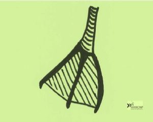
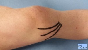
Just below the knee on the inside of the leg there is a structure called pes anserinus, which literally means goose’s foot! As 3 tendons come together at this point, it resembles the appearance of a goose’s foot.

Illustration of webbed goose foot
The 3 tendons that form the pes anserinus – sartorius, gracilis & semitendinosus.


There are usually underlying biomechanical issues, which means that excessive strain is being placed on the tendons and attachment area of the goose’s foot.

Pes anserinus injury is highly painful and usually results in having to cease running or walking and even causes a limp.

The good news is that alleviating the pain can be achieved by:

  • using anti-inflammatory measures
  • taking the strain off the injured tendons (e.g. taping)
  • limiting activity levels
  • soft tissue techniques

Whilst getting rid of the pain is all well and good, the most important thing for those with pes anserinus issues is to address the biomechanical issues underlying the condition and allow return to physical activity.

This requires a thorough assessment with an experienced physiotherapist with a sound understanding of lower limb biomechanics. Altered biomechanics can be addressed in a number of ways including:

  • specific strengthening exercises
  • targeted stretching
  • changes in footwear or orthotics

If you think you may may be having issues with your goose’s foot, we can help you get out of pain, get back exercising and improve your biomechanics. To make an appointment with one of our experienced physios phone 8850 7770 or click here.

Do you ever shake your hands to rid pain or regain sensation in your hands?

  • Do you find yourself waking at night with hand pain or numbness?
  • Are you frequently shaking your hands due to lack of sensation?
  • Do you get shooting pain up your forearm?

If so, you may have Carpal Tunnel Syndrome.

Carpal Tunnel Syndrome is more common than you may think and we’ve been seeing it (to varying extents) in a large proportion of our patients. It can also be a highly debilitating condition impacting many aspects of daily life.

What is Carpal Tunnel Syndrome?

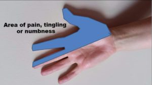
Carpal Tunnel Syndrome is damage or irritation to the median nerve as it passes through the carpal tunnel (a ring of bones in the wrist). The result of damage to the nerve is commonly pain, pins & needles or numbness in the hand (typically the palm side of the hand, including the thumb, index and middle fingers). These symptoms can occur at any time, although they are often worse at night with people frequently waking with a dead hand sensation as well as having severely disrupted sleep.

If Carpal Tunnel Syndrome is left untreated, it can result in:

  • Persistent pain, tingling and/or numbness in the hand,
  • Weakness in the hand, which may lead to functional impairments (including reduced grip strength and hand dexterity),
  • Additional nerve damage further up the arm and even in the neck,
  • Permanent damage to the median nerve.

Those with Carpal Tunnel Syndrome will tell you that they will do anything to get rid out it. Why wouldn’t you want a pain-free, fully functioning hand with normal sensation? Or perhaps an uninterrupted night of sleep, the ability to open jars again or not having to shake your hand to “wake it up”?

Tailored physiotherapy programs can treat the symptoms of Carpal Tunnel Syndrome that you may have. Physiotherapy treatments for Carpal Tunnel Syndrome include:

  • Splinting, which involves wearing an appropriate brace to ease the strain and reduce the irritation on the median nerve.
  • Nerve gliding exercises, which are specific exercises to get your median nerve moving well through the carpal tunnel as it often gets “stuck” when it is irritated.
  • Wrist and forearm stretching and strengthening exercises.
  • Addressing postural concerns and neck issues which can be associated with Carpal Tunnel Syndrome.

Our physiotherapists are well-versed in identifying and assessing for Carpal Tunnel Syndrome, as well as differentiating this from other similar conditions.

If you are waking in the night with hand pain or think you may be experiencing Carpal Tunnel Syndrome, a consultation with one of our physios will help put a plan together to put an end to it. Phone 8850 7770 or click here and we’ll help you get on top of it ASAP.